
Chapter 3: Nutrients in the Body
3.5 Small Intestine
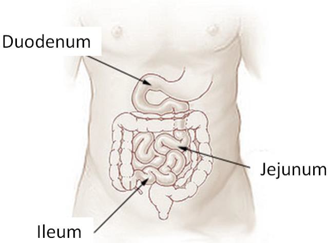
The small intestine is the primary site of digestion. It is divided into three sections: the duodenum, jejunum, and ileum (shown below). After leaving the stomach, the first part of the small intestine that chyme will encounter is the duodenum.
The small intestine consists of many layers, which can be seen in the cross section in Figure 3.52 below.
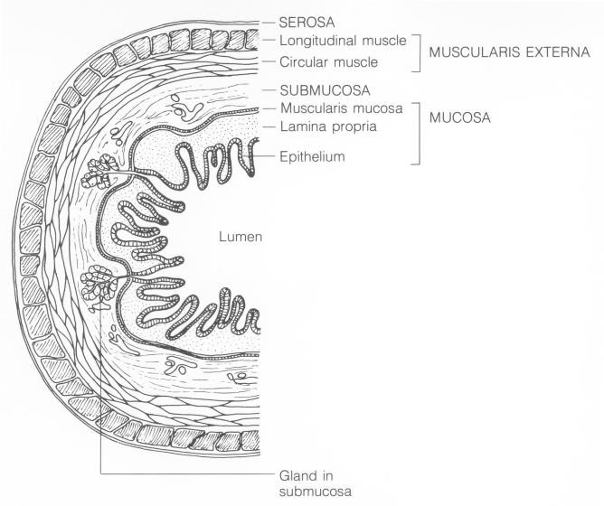
Examining these layers more closely, we are going to focus on the lining of the small intestine, known as the epithelium (see Figure 3.52 above), which comes into contact with the chyme and is responsible for absorption. The lumen is the name of the cavity that is considered “outside the body” that chyme moves through.
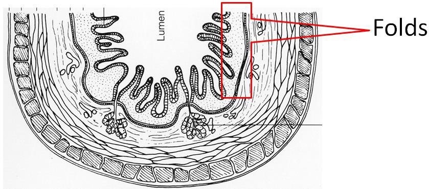
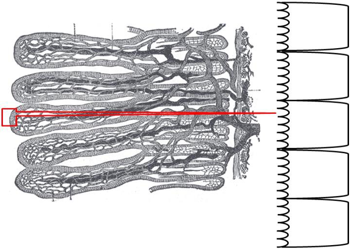
The organization of the small intestine is in such a way that it contains circular folds and finger- like projections known as villi. The folds and villi are shown in the next few figures.
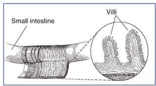
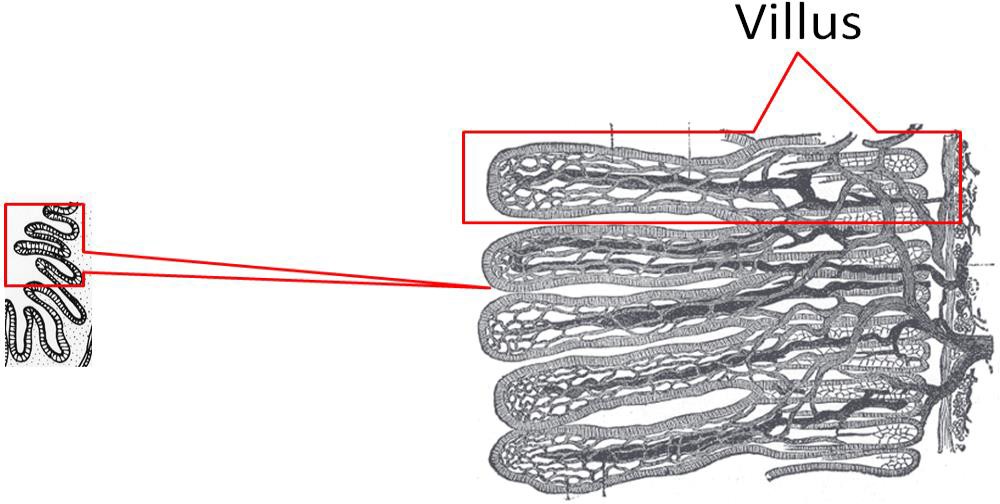
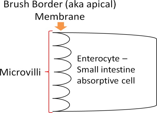
If we were to zoom in even closer, we would be able to see that enterocytes (small intestine absorptive cells; a.k.a brush border cells) line villi as shown below. This layer is referred to as the mucosa, and is composed primarily of simple columnar epithelium.
The side, or membrane, of the enterocyte that faces the lumen is not smooth either. It is lined with microvilli, and is known as the brush border membrane, as shown below.
Together these features (folds + villi + microvilli) increase the surface area ~600 times versus if it was a smooth tube[1]. (Note: the symbol ~ is used in place of the word “approximately.” You will see it used other places in this text as well.) More surface area leads to more contact between the chyme and the enterocytes, and thus, increased absorption.
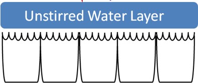
Finally, the surface of the cells on the microvilli are covered with proteins, which helps to catch a molecule-thin layer of water within itself. This layer, called the “unstirred water layer,” has a number of functions in absorption of nutrients, and will have a direct impact on fat absorption as we will see later[2].
- Byrd-Bredbenner C, Moe G, Beshgetoor D, Berning J. (2009) Wardlaw's Perspectives in Nutrition. New York, NY: McGraw-Hill. ↵
- http://www.newworldencyclopedia.org/entry/Small_intestine ↵
Microvilli are microscopic cell extensions that increase the surface area of a cell. They do not move. An example is the brush border membrane on intestinal cells.
