
Chapter 3: Nutrients in the Body
3.8 Absorption and Circulation Overview
The term absorption can have a number of different meanings. In nutrition, absorption means that a compound is taken in from the lumen of the digestive tract into the bloodstream for circulation throughout the body. This process is sometimes divided into two steps: uptake (compounds being moved from the lumen into the enterocyte) and absorption (a compound being moved from the enterocyte to the bloodstream). Under most circumstances, compounds that are taken up into the enterocytes will then be absorbed into the bloodstream.
What’s Actually Absorbed
We know that digestion breaks large macronutrient molecules into smaller molecules. It is these smaller molecules that are absorbed into the bloodstream. The figure below shows what is actually taken up into the enterocyte. We will go into more detail about how these small molecules are made when we discuss digestion of specific macronutrients in chapters 5, 6, and 7.
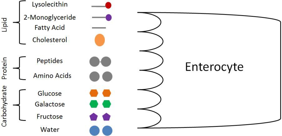
From lipids, we have lysolecithin (from phospholipids), 2-monoglyceride (from triglycerides), fatty acids, and cholesterol. From protein, there are small peptides (di- and tripeptides) and amino acids. From carbohydrates, only the monosaccharides glucose, galactose, and fructose will be taken up. The other macronutrient, water, has not been discussed so far because it does not undergo digestion.
Vitamins and minerals are also taken up through enterocytes.
The Plasma Membrane
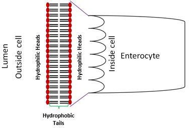
In order to be taken up by the enterocytes, these compounds must now cross the cell (plasma) membrane, which is a phospholipid bilayer. In the cell membrane, the hydrophilic heads of the phospholipids point into the lumen of the GI tract, as well as towards the interior of the cell, while the hydrophobic tails are on the interior of the plasma membrane. This is depicted in the diagram below.
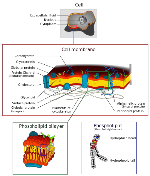
In addition to phospholipids, the cell membrane also contains proteins, cholesterol, and carbohydrates in addition to the phospholipids. Membrane proteins, such as channels, pumps, pores, and carriers are important for the transport of some compounds across the cell membrane. Figure 3.83 and two videos below do a nice job of illustrating the components of the cell membrane.
Transport Across the Plasma Membrane
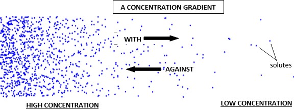
There are a number of different transport mechanisms utilized by your body for the uptake of nutrients into cells, and absorption into the bloodstream. These mechanisms can be classified as being either passive transport or active transport. The difference between the two types of transport is whether energy is required, and whether they move with or against a concentration gradient. Passive transport does not require energy and moves with a concentration gradient (high to low concentration). Active transport requires energy to move against the concentration gradient (low to high concentration).
A concentration gradient is a result of an unequal distribution of solutes within a solution. In a solution, a solute is dissolved in the solvent. For example, in salt water, the solute is salt and water is the solvent. The more solute a region has, the higher the its concentration, while the less solute a region has, the lower the its concentration. Moving with the gradient is moving from a region of higher concentration to an area of lower concentration. Moving against the gradient is moving from an area of lower concentration to an area of higher concentration.
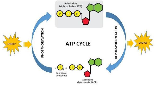
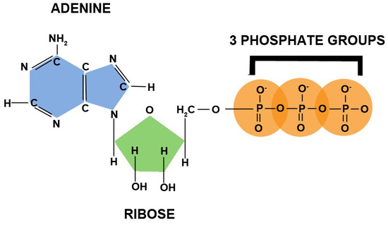
The energy for active transport is provided by adenosine triphosphate (ATP), which is the energy currency in the body. Tri- means three, thus ATP is adenosine (composed of an adenine and a ribose) with three phosphate groups bonded to it, as shown below.

Phosphorylation is the formation of a phosphate bond. Dephosphorylation is the removal of a phosphate bond. Phosphorylation is an anabolic process that requires energy.
Dephosphorylation is a catabolic process that releases energy. Thus, energy is required to add phosphates to ATP, while energy is released through removing phosphates from ATP. Figure 3.86 depicts this process.
Passive Transport Mechanisms
There are three forms of passive transport involved in uptake and absorption of nutrients in the body:
- Simple Diffusion
- Osmosis
- Facilitated Diffusion
Simple Diffusion
Simple diffusion is the movement of solutes from an area of higher concentration to an area of lower concentration (with the concentration gradient) without the help of a protein, as shown in Figure 3.87.
Osmosis
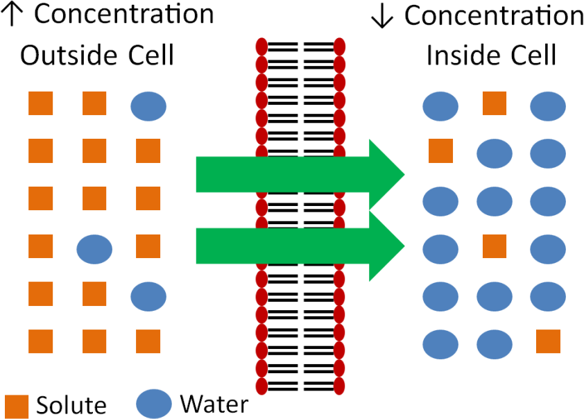
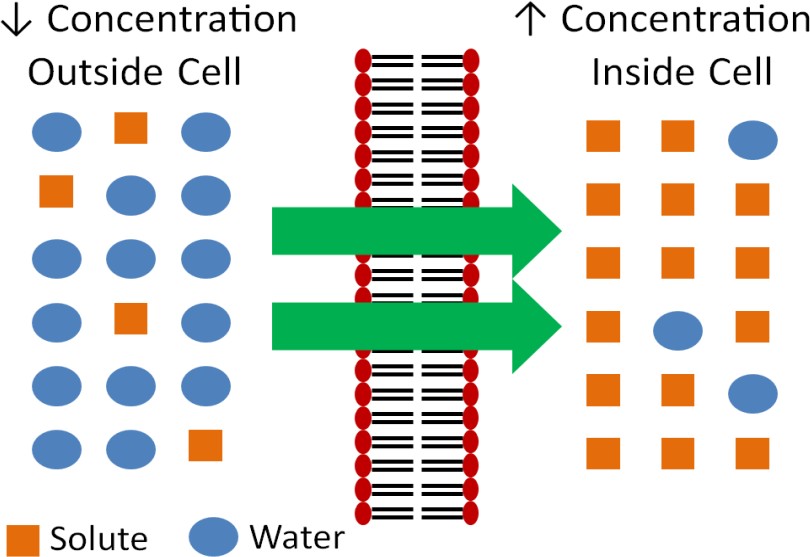
Osmosis is similar to simple diffusion, but water moves instead of solutes. In osmosis water molecules move from an area of lower solute concentration to an area of higher solute concentration as shown below. The effect of this movement is to dilute the area of higher concentration.
The following videos do a nice job of illustrating osmosis.
Another example illustrating osmosis is the red blood cells in different solutions shown below.
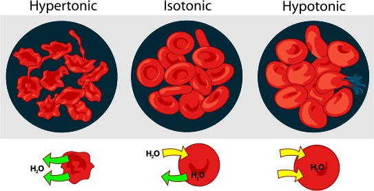
We will consider the simple example of salt as the solute. If the solution is hypertonic, that means that there is a greater concentration of salt outside (extracellular) the red blood cells than within them (intracellular). Water will then move out of the red blood cells to the area of higher salt concentration, resulting in the shriveled red blood cells depicted. Isotonic means that there is no difference between concentrations. There is an equal exchange of water between intracellular and extracellular fluids. Thus, the cells are normal, functioning red blood cells. A hypotonic solution contains a lower extracellular concentration of salt than the red blood cell intracellular fluid. As a result, water enters the red blood cells, possibly causing them to burst.
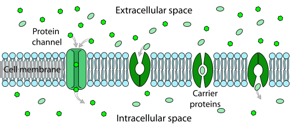
Facilitated Diffusion
The last form of passive transport is similar to diffusion in that it also moves with the concentration gradient (higher concentration to lower concentration). While it requires no energy, it does require a carrier protein to transport the solute across the membrane. Figure 3.810 and the video link below do a nice job of illustrating facilitated diffusion.
Video Link: Facilitated Diffusion (0:27)
Active Transport Mechanisms
There are two forms of active transport:
- Active Carrier Transport
- Endocytosis
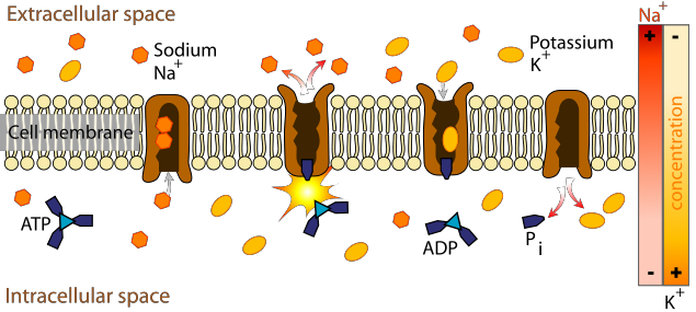
Active Carrier Transport
Active carrier transport (sometimes referred to as secondary active transport) is similar to facilitated diffusion in that it utilizes a protein carrier. However, energy is also required to move compounds against their concentration gradient (lower to higher concentration). Figure 3.811 and the video below do a nice job of illustrating active carrier transport.
Video Link: Active Transport (0:21)
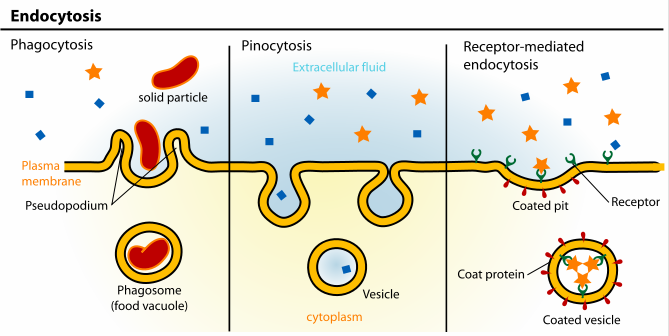
Endocytosis
Endocytosis is the engulfing of particles, or fluids, to be taken up into the cell. If a particle is endocytosed, this process is referred to as phagocytosis. If a fluid is endocytosed, this process is referred to as pinocytosis. Whenever a receptor located on the membrane is used to assist in engulfing an extracellular component, it is known as receptor mediated endocytosis. These processes are shown in Figure 3.812.
The following video does a really nice job of showing how endocytosis occurs.
Video Link: Endocytosis (0:35)
Circulation
There are two types of vessels that take up absorbed nutrients once they cross the intestinal wall: capillaries and lacteals. Capillaries are part of the circulatory system, the smallest blood vessels in your body. Lacteals are part of the lymphatic system, they are filled with tissue fluid called lymph. Most absorbed nutrients are taken up into the capillaries. The lacteals take up fat droplets that are too big to enter the capillaries. (This makes the lymph in the lacteals a milky white, which is why they’re called lacteals!)
The diagram below shows two individual villi. The lacteals are in green and the capillaries in red and blue. Each villus in the small intestine has a lacteal and several capillaries running up the middle of it, perfectly positioned to pick up all of the nutrients absorbed through the intestinal cells (red arrows).
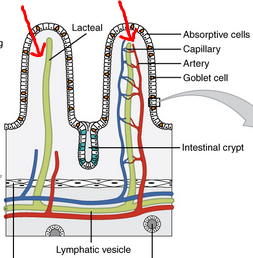
Nutrients that are absorbed into capillaries are swept directly to the liver for processing. Fats taken up by the lacteals bypass the liver initially and travel through the lymphatic system. This fluid is eventually returned to the bloodstream just before blood returns to the heart.
Summary of Nutrient Absorption
The figure below provides a summary of nutrient absorption. It shows the digestion of large molecules in the lumen of the small intestine and smaller molecules (products of digestion) being taken up across enterocytes. At the bottom of the figure, single sugars and amino acids enter capillaries (in red) for circulation to the liver, and lipids enter the lacteals (in yellow), for eventual entry into the bloodstream.
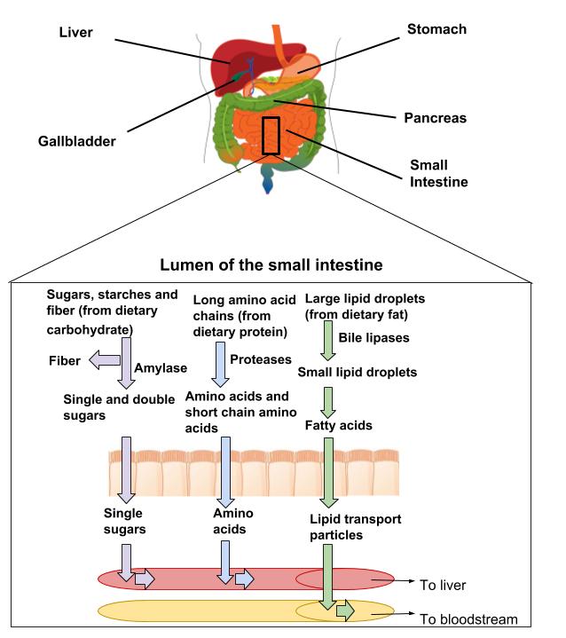
We will go into greater detail on digestion and absorption of specific nutrients in later chapters.
Absorption is the process of getting nutrients from the digestive tract into the blood or lymph.
Enterocytes are the cells that line the villi of the small intestine and absorb nutrients. (also known as enteric cells or brush border cells).
Amino acids are the building blocks of proteins, simple subunits composed of carbon, oxygen, hydrogen, and nitrogen.
Monosaccharides are the simplest of all sugars and are the building blocks of all carbohydrates. The monosaccharides are glucose, fructose, and galactose.
Vitamins are organic compounds that must be taken in from the diet.
Minerals are solid inorganic substances that form crystals and are classified depending on how much of them we need. Trace minerals such as zinc, iron, or iodine are only required in a few milligrams or less per day. Major minerals such as calcium, sodium, and potassium are required in hundreds of milligrams per day.
Concentration gradient is a result of an unequal distribution of solutes within a solution.
Passive transport is the transport of molecules across the cell membrane without the use of ATP.
Active transport requires energy to move against the concentration gradient (low to high concentration).
Solutes are substances dissolved in a fluid (the solvent).
Villi (singular: villus) are finger-shaped projections from the inner wall of the small intestine.
